What do we see in the air we breathe?
Dec. 19, 2022
A first look at our filters with microscopy.
An earlier blog discussed why we’re using filters to capture Melbourne’s air. You also asked us what else is on the filter, other than pollen.
Our friends at the University of Melbourne Materials Characterisation and Fabrication Platform (MCFP) have generously offered to take a deep dive look at one of the filters using some of the advanced equipment they have that’s well-suited to identifying the materials on it.
For an initial look, Dr Anders Barlow used a high performance “optical” microscope that uses a combination of intense lighting with very high-quality glass lenses to capture and focus light onto a digital camera (Figure 1).
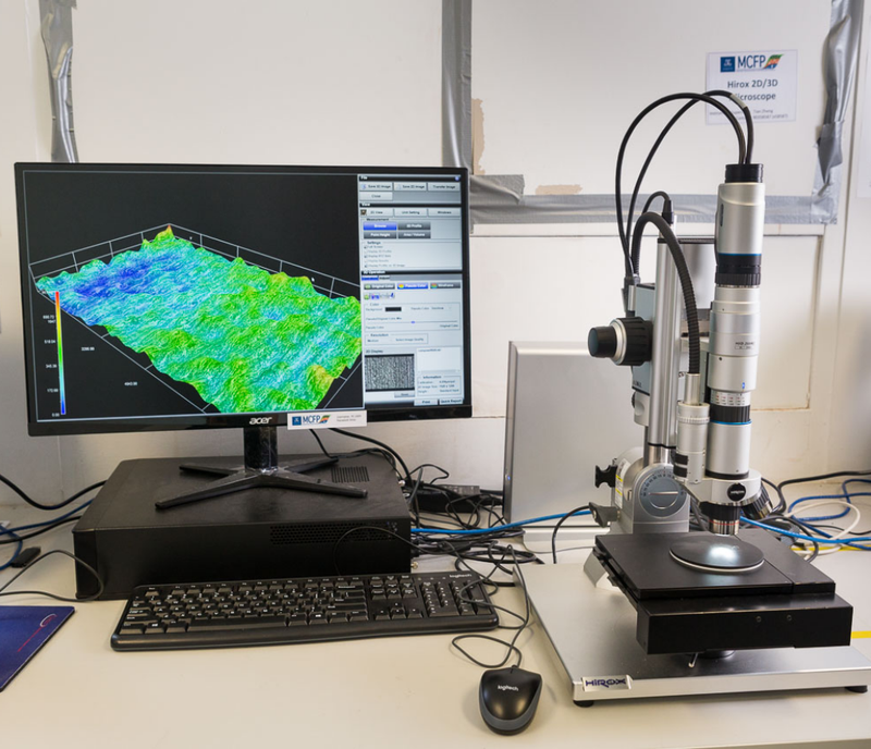
Figure 1: High-performance Hirox RH-2000 microscope.
Using this microscope we can see (Figure 2) that the clean filter is almost featureless at this scale, you can just barely make out the tiny strands of the filter material with this type of microscope.
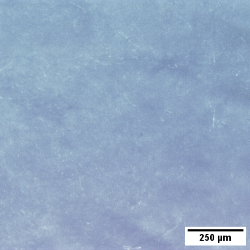
Figure 2: Image of clean filter taken with a Hirox RH-2000 microscope.
In contrast on the used filter (Figure 3) you can see a lot of “material” has been caught by the mesh and we see a number of larger embedded particles of different shapes, colours and sizes.
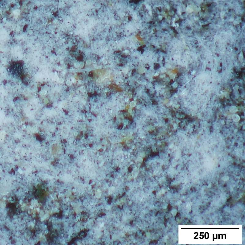
Figure 3: Image of a filter after it has been collecting material in Melbournes air for 7 days, taken with a Hirox RH-2000 microscope.
Zooming a little further in (Figure 4) it is somewhat easy to identify pollen grains on the filter because they are relatively “large” with a diameter of about 30-50 micrometres (0.03 to 0.05 mm), but to see anything at higher magnification we’ll need to use a different approach because optical techniques, i.e. those that use visible light, are limited to a resolution of about 1 micron (one-thousandth of a millimetre, 0.001 mm) or a bit less in size. For comparison, human hair is on average about 10-50 microns thick.
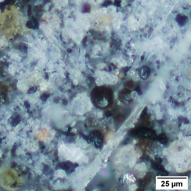
Figure 4: A higher magnification image of a filter after it has been collecting material in Melbournes air for 7 days, taken with a Hirox RH-2000 microscope.
To see things at even higher magnifications we need to switch to a microscope that uses beams of charged particles to illuminate materials. Commonly these are electron beams or ion beams, and the instruments are called a scanning electron microscopes (SEM) or helium ion microscopes (HIM). In our next blog post, we’ll show you what the filter looks like using these sorts of Microscopes.
© 2010- AirHealth Pty Ltd. All rights reserved.
This information is copyrighted (Terms & conditions). Apart from any use as permitted under the Copyright Act 1968, no part may be reproduced by any process without prior written permission. This includes all pollen information and forecasts.